ADDRESS:
CTLAB
Central European Institute of Technology
Purkynova 123, 612 00 Brno
Czech Republic
+420 541 142 875
VAT: CZ00216305


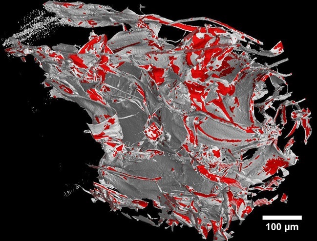
The 3D imaging of cell using Rigaku Nano3DX
Our new publication in Journal of Microscopy is foccused on 3D imaging of mesenchymal stem cells on porous scaffolds using Rigaku Nano3DX with a voxel resolution 540 nm. To evaluate the potential of nano-CT, a comparison measurement was done using scanning electron microscopy (SEM) combined with energy dispersive X-ray analysis (EDX). The proposed method will help to understand better the behaviour of cells while interacting with three-dimensional biomaterials. This is crucial for clinical tissue engineering applications in order to limit the risk of uncontrolled cell growth and potentially tumour formation. Full paper is available here: https://onlinelibrary.wiley.com/doi/pdf/10.1111/jmi.12771.