
CEITEC CTLAB
Central European Institute of Technology
Purkynova 123, 612 00 Brno
Czech Republic
+420 541 142 875
VAT: CZ00216305


Our new article in Scientific Reports
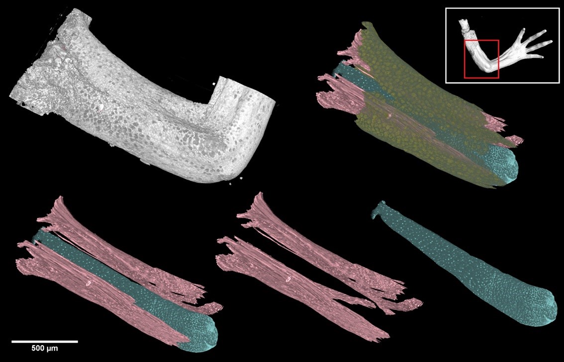
It is generally known that if the salamander loses its limb, it will be renewed. The regeneration capability of salamanders and other amphibians was described long time ago. But, what is not known is how this mechanism occurs in nature.
An important step in clarifying the regeneration mechanism is to observe the geometry of skeletal and muscle structures during the growth and development of salamander. Markéta Tesařová, PhD student from our Laboratory of X-ray micro nad nano Computed Tomography at CEITEC BUT has been working on this topic together with biologists from Karolinska Institutet for more than two years. Her research has been focused on visualization, quantification and analysis of different structures of salamander with cellular resolution in three-dimensions. The results of her investigation was recently published in prestigious Scientific Reports journal (Nature Publishing Group, IF 4.6).
In this work, synchrotron X-ray computed microtomography was exploited for a quantitative analysis of the 3D-cell distribution in tissues of a developing salamander (Pleurodeles waltl) limb – a key model organism for vertebrate regeneration studies. Using the tomographic approach, cell polarity was studied in relation to the developing joints of the salamander model. Visualization of these structures in 3D is the first step to a chance to cure injuries or illnesses in which defined cell type dies in the body.
Full paper is available here: https://www.nature.com/articles/s41598-018-32459-2#article-info.